Tennis Elbow – Part 2

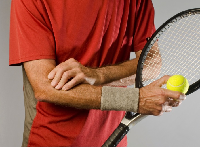
In the first part of this article series, we discussed less common causes of lateral elbow pain that may present similarly to tennis elbow. Tennis elbow is, however, still by far the most common cause of pain in this area. In this second part of the series we discuss some common treatment approaches for this condition.

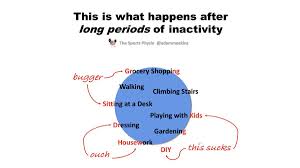
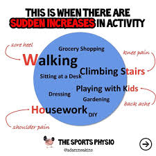
Tennis elbow, or lateral epicondalgia/lateral elbow tendinopathy, occurs due to an overload of the tendons of the wrist extensor muscles that attach to the forearm close to the elbow joint. The condition is thought to arise secondary to some type of increase in loading, either acutely (a single bout of excessive or unaccustomed activity) or chronically (increased activity over a prolonged period of time). It is also thought that there is some degree of degeneration or natural wear to the tendons that may predispose an individual to the condition, however we do also frequently see these structural changes in people without elbow pain. 

There may also be a biomechanical element to the issue. Tightness or weakness in other areas of the body may lead to increased pressure being put on the elbow. In the case of sporting actions that have led to the problem, there may also be technique issues that can contribute to the increased loading on the elbow. 

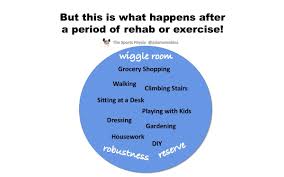
As with all tendon issues, the key to recovery is proper management of load. This may be a temporary change in activity levels, modifications to movement to decrease load, or even taping and bracing strategies to offload the injured area. The other side of the load management equation, however, involves specific exercise to build up the capacity to tolerate loading. As the movements most commonly affected by tennis elbow are gripping and wrist extension, our specific exercises usually involve loading of one or both of these movements. The key is finding the right amount of loading to cause an adaption, improve capacity and decrease pain with these movements, whilst not being excessive as to aggravate the condition. Based on the outcomes of a physical examination including a symptom modification procedure performed by a physiotherapist or osteopath, most patients will respond to a particular type of glide to change the pain with these movements and allow initial loading to occur. Below are two examples of exercises involving this symptom modification procedure. 

From here we can progress the rehabilitation in a number of different ways depending on functional goals of the patients, as well as other factors such as age. The goal with any patient is to progress the loading on the elbow to the stage where it can tolerate the demands of the patient’s lifestyle and activity levels. Below are some examples of exercises that may be used to progress the loading on the elbow. 


As discussed earlier in the article, weakness and tightness in other areas of the body may also be influencing the loading on the elbow, and so the exercise program should also address and optimise these factors where possible. 

There are some cases where tennis elbow fails to respond to conservative management, even after a long period of time and some patients will opt for slightly more invasive methods. The most commonly utilised of these are shockwave and corticosteroid injection. Whilst having some evidence of positive effect in other joints, the evidence for shockwave in the treatment of tennis elbow is mixed, and its use doesn’t guarantee a quicker or more complete recovery. 

Corticosteroid injection has been shown to have some short-term benefit, however the long-term prognosis isn’t better than that of a more conservative approach and these injections do carry with them the risk of tendon rupture. Hence, these modalities should only be used once all other measures have been exhausted and with a patient who fully understands their risks. 

The management of tennis elbow can be a long and arduous journey for the patient, however with an individualised approach and closely monitored rehabilitation program, chances of return to previous activity levels are high. 

If you or someone you know is suffering from lateral elbow pain we are here to help! Contact us now and let us help get you back on track.

Sam head shot

Sam Gilbert is the co-founder and clinical director of Club 360. He has a bachelor’s degree in physiotherapy and a master’s degree in exercise science.

Tennis Elbow – Part 1

Tennis Elbow – Part 1 

Tennis elbow (also commonly referred to as lateral epicondalgia, lateral elbow pain, lateral elbow tendinopathy, lateral epicondylitis) is a very common condition in musculoskeletal practice. This first part of a two part series will look at some other conditions that present very similarly to tennis elbow. 

Whilst pain on the lateral (outside) aspect of the elbow is commonly originating from the tendons of the wrist extensor muscles (the classic “tennis elbow”), there are a number of other conditions that may present as pain in the area. Whilst traditional tennis elbow is notoriously slow to respond to treatment interventions, another reason for prolonged recovery time is inaccurate diagnosis, and subsequently an inappropriate treatment plan. 

Elbow joint pain

The first area that may mimic tennis elbow is the elbow joint itself. Joint pain should be suspected particularly in younger patients. Tennis elbow is a condition that typically affects people between 35 and 60. For younger, especially very active people, it is likely that there is joint involvement. Another clue is if the pain is worse in elbow flexion (with the elbow bent) as typical tennis elbow is worse with the elbow straight. 

Structures around the elbow joint that are thought to cause pain are the ligaments (particularly the lateral collateral ligament, or LCL), the plica (a fold of joint tissue that can become squeezed between the bones of the upper arm and the forearm), the cartilage of the joint, and the bursa (a sack of fluid that acts as a shock absorbing structure). For patients with a history of acute elbow injury, or a history of high force through work or sport, osteoarthritis may develop in the elbow joint and cause pain in the lateral aspect. 

In children, a condition called osteochodrosis dissecans may cause pain and loss of function, where pieces of bone in the joint start to separate from the main bone due to a lack of blood supply. As tennis elbow is quite rare in this population, children suffering ongoing elbow pain should undergo appropriate imaging in order to make an accurate diagnosis, especially due to the serious outcomes associated with mismanagement. 

Nerve pain

It is quite common for the radial nerve to cause pain in the outside of the elbow, and this nerve may become entrapped or inflamed at a number of different sites, including the neck and shoulder region, the radial tunnel at the elbow, and within some of the muscles around the elbow and forearm, in particular the supinator muscle. Sometimes pain of neural origin will be accompanied by other neural symptoms such as numbness, weakness, and pins and needles. The type of neural symptoms, including their location and the nature, will differ depending on the area of entrapment, and as such accurate diagnosis is important. Some of the treatment modalities sometimes used to address tennis elbow, such as bracing and massage of the forearm muscles, will actually increase the pressure around the sensitised nerve and increase pain.

Systemic Conditions

Systemic inflammatory conditions such as spondyloarthropathies may also cause pain in the area. Patients with pain in other joints, symptoms related to the eyes, skin or digestive system, or a personal or family history of systemic inflammatory disorders should undergo appropriate testing to rule this out as a source of elbow pain. 

Summary

The management for any of the above conditions will likely be very different to that of tennis elbow, and as such it is imperative that an accurate diagnosis is made. If you or someone you know are experiencing lateral elbow pain, please contact your trusted medical professional to have the injury properly diagnosed to optimise your chances of a positive outcome.


Next week in part 2 of this series we discuss possible management options for tennis elbow.

Sam head shot

Sam Gilbert is the co-founder and clinical director of Club 360. He has a bachelor’s degree in physiotherapy and a master’s degree in exercise science.


Heel the World

What is Plantar Fasciopathy (“Plantar Fasciitis”)

Plantar (bottom of the foot) heel pain is something we see very often in clinic, and tends to be extremely frustrating and debilitating for the sufferer. 

Pain in this area is typically due to an increase in sensitisation around the insertion of the plantar fascia, a thick band of collagen that runs between the heel and the balls of the feet. The plantar fascia has an important role in the biomechanics of the foot, providing structure to the arch and aiding in propulsion during the gait cycle. When excessive load is placed through the feet, either by prolonged standing or walking, increased volume of running, poor biomechanics, or even increased bodyweight, the structure can become overloaded and start producing pain. 

download-4

Patients will often complain of pain particularly upon the first few steps in the morning. The pain may ease with activity, only to return again following cessation.

This condition was for a long time referred to as “plantar fasciitis”, as the belief was that the pain was caused by a persistent inflammation of the fascia, and treatment focused on stretching, orthotic prescription and analgesic modalities such as ice. We now understand the nature of this pathology to be more like that of persistent tendon pain, whereby the issue is less of an inflammatory one and more related to change in the structure of the collagen and general increase in sensitivity. Therefore the medical community as a whole has opted for more appropriate terms such as plantar fasciopathy or plantar heel pain. Many patients will have gone through medical investigations including x-ray, and may have been identified as having a calcaneal bone spur. This was for a long time thought to be the cause of pain in the heel, however we now understand that whilst the prevalence of these spurs is much higher in those individuals with a history of heel pain, it isn’t the bone spur itself that causes the pain, and resolution of symptoms is likely without addressing the spur directly. 

The treatment for plantar fasciopathy begins with addressing the likely causative factors. These may be changes in activity load, biomechanics (i.e. running gate, relative weakness/tightness of muscles etc.), changes in body weight etc. For active individuals this may require a period of decreased exercise, or temporarily switching to alternative exercise modalities. Another important aspect of the initial management is education regarding the nature of the condition, and setting realistic time frames for recovery, as plantar fasciopathy is notorious for taking a long time to resolve. Once a baseline of activity modification adequate for improvement in symptoms has been identified, load can be gradually increased based on the pain response. 

In the early stages, rubber heel cupping, taping, and orthotics may aid in settling symptoms. As early as possible, strengthening exercises should be commenced to improve the strength, endurance and power of the local muscles, as well as improving the load tolerance capacity of the plantar fascia itself, which will decrease its likelihood of being overloaded and causing pain. These should be progressed to functional conditioning work specific to the activity that the individual is aiming to return to. 

Other Causes of Heel Pain 

In addition to plantar fasciopathy, there are several other conditions that may cause pain in the base of the heel, including bruising of the fat pad, stress fracture of the heel, systemic rheumatological conditions, as well as entrapment of nerves around the foot and ankle. As these conditions can all mimic the typical presentation of plantar fasciopathy, it is important that the condition is evaluated by an experienced orthopedic medical professional, as the management for these conditions will differ, and incorrect treatment could cause a delay in resolution or even a worsening of the condition. 

If you are suffering heel pain don’t hesitate to get in contact with us so that we can get you on the path to recovery as soon as possible. 

Sam Gilbert is the co-founder and clinical director of Club 360. He has a bachelor’s degree in physiotherapy and a master’s degree in exercise science.

Sam head shot

Safely Returning to Exercise after Lockdown 

Whilst the COVID-19 pandemic is still wreaking havoc across the globe, some parts of the world, including Japan and Australia, are beginning to ease restrictions and reintroduce certain services. These changes will likely allow individuals to partially or fully resume their normal sports and fitness practices. Current literature regarding secondary health consequences of the COVID-19 have so far been focused around inactivity(1), and whilst the health benefits of resuming physical activities are obvious, there are some important considerations when it comes to avoiding overuse injury.

All types of physical activity impose stress on both the tissues (muscles, bones, ligaments, tendons, etc.) as well as the multiple systems (neural, cardiovascular endocrine, etc.) of the body. It is the adaptation that follows this stress that allows us to improve different aspects of our fitness. In order for these adaptations to occur, there needs to be the correct amount of training stimulus. If this stimulus is too small, the exercise modality will be ineffective. If it is too large, the tissues and systems may become compromised, and this is where we can sometimes get into trouble. When we have gone for a certain period of time without performing an activity, our body’s optimal loading zone changes, so that the correct amount of stimulus required to cause adaptation without overloading the system is now less. If we then go back to performing the same type, volume and intensity of exercise that we were doing before, we may risk sustaining an overuse injury. The diagrams below, courtesy of UK physiotherapist Adam Meakins, illustrate that optimal zone of activity and how going outside this zone can start to cause problems. 

images-1images-3

imagesimages-2

Sports performance professionals have for a long time focused on workload management. Runners will no doubt be familiar with the 10% rule, and researchers in recent times have discussed the concept of the acute:chronic workload ratio, whereby increases in current training load outside a certain range compared to your average workload may increase your risk of overuse injury. The first to explore this concept were Gabbett and colleagues, who found that a training load outside a 85-135% range in comparison to the average load over the previous 4 weeks to significantly increase injury rates(2). What is particularly interesting is that the injuries that were thought to occur as a result of this change in training load in some cases didn’t eventuate for 1-2 months after the load change(3).

Many individuals who have been unable to participate in their regular physical activities for a period of time are likely to throw themselves back in at the same volume and intensity as prior to the break, or maybe even with greater vigour due to having missed the enjoyment and

 social interaction of the activity, or possibly even due to feelings of guilt and concern over being inactive for so long. When one returns to sport or exercise after injury or severe illness, it is somewhat inferred that this return needs to be performed as a gradual process, however in the absence of physical impairment, a more-is-better approach is too often adopted.

Even for those who have continued exercising, we need to consider the impact of varying exercise modalities on injury risk. If, for example, a person who is a regular swimmer starts running due to their pool being closed, then resumes a high-load of swimming as soon as the gym reopens, the unaccustomed load may put excessive stress on the upper limbs. 

Here are some simple tips to decrease the likelihood of post-lockdown return to activity leading to an overuse injury. 

  • Rather than jumping straight back into your previous exercise load, think about return to activity more as a medium-term process. Think 4 weeks ahead and plan on building back to your previous load within this time. Then start at approximately 50% of this load and build up gradually.
  • Try to build up the volume quicker than the intensity. If performing resistance training try to stay further away from failure with each set. If performing cardiovascular training try to perform slightly more steady state exercise and build back up to more interval work.
  • Spend more of your gym time warming up and cooling down. That way you are still getting your gym time and movement in, but not working quite so hard the whole time. Mobility is likely to be compromised during periods of inactivity, so spending more time on mobility interventions is likely to lead to better long-term progress.
  • Also consider the total load on the system. Things such as increased stress, decreased sleep, dietary changes, etc., all contribute to total systemic load. If there are other changes to your lifestyle involved with a return to the office, consider a slower return to exercise to avoid system overload.

Remember that in order to make fitness gains we want the minimal effective dose, that way can continue to build up our training load over time, and still progress. If we do too much too quickly, we may hit a ceiling too quickly and find it more difficult to progress. This is the case not only when beginning a certain type of exercise but also after a long lay-off. 

The whole world is looking forward to a gradual return to normally, and for many, resuming regular exercise habits constitutes a big part of this. We always maintain that exercise is like medicine, and needs to be dosed properly for optimal outcomes. Just applying a little patience and intelligence to your exercise programming will pay off in the long run and allow you to get back to the activities you love, safely and healthily.

References 

  1. Lippi, G., Henry, B. M., Bovo, C. & Sanchis-Gomar, F. Health risks and potential remedies during prolonged lockdowns for coronavirus disease 2019 (COVID-19). Diagnosis 1, (2020).
  2. Hulin, B. T., Gabbett, T. J., Lawson, D. W., Caputi, P. & Sampson, J. A. The acute: chronic workload ratio predicts injury: high chronic workload may decrease injury risk in elite rugby league players. Br. J. Sports Med. bjsports–2015 (2015).
  3. Rogalski, B., Dawson, B., Heasman, J. & Gabbett, T. J. Training and game loads and injury risk in elite Australian footballers. J. Sci. Med. Sport 16, 499–503 (2013).

Sam Gilbert is the co-founder and clinical director of Club 360. He has a bachelor’s degree in physiotherapy and a master’s degree in exercise science.

Sam head shot

The Right Leg’s Connected to the … Left Leg?

Rehabilitation of any injury or condition is a multifaceted process that requires attention to every aspect related to a successful recovery. One of the factors often overlooked is the maintenance of conditioning on the non-affected side. What many individuals, including coaches and clinicians, often fail to appreciate is the fact that training the non-injured side of the body can lead to a faster and more complete recovery, through a process known as cross education.


Cross education (also referred to as “cross transfer”, “interlateral transfer”, “cross limb transfer”, “contralateral training effect”, and “interlimb transfer”) was first referred to in the literature in 18941 and has been thoroughly investigated in over 20 clinical trials. It is basically the phenomenon whereby training one side of the body leads to strength changes on the opposite side.


Broadly speaking, the two main mechanisms by which force production capacity can be improved are 1) changes in the muscular tissue and 2) changes in the nervous system, or put more simply, improvements in the hard-wiring to the muscles. These nervous system changes take place at numerous different sites, including the brain itself. As you are probably aware, there are two hemispheres of the brain. These two hemispheres are linked by numerous structures, in particular an important bridge called the corpus callosum. These structures allow for communication between the two hemispheres, and for the benefits of training stress to be shared between both sides of the body. Essentially the brain is not smart enough to figure out where the training stimulus is coming from and as a result makes both sides of the body stronger.


Studies have shown strength gains in the non-trained limb of up to 77% of the trained side2. Amongst the research siting evidence of it’s effectiveness the most relevant to rehabilitation professionals are the three studies showing increased strength and a maintenance of muscle mass on the opposite side even when the limb was immobilised3,4,5. Whilst these studies haven’t been repeated on injured individuals, it does suggest that if we can maintain strength training on the opposite side we can potential ward off some of the strength and muscle mass losses that inevitably occur following injury, and therefore optimise the rehabilitation process.


There are other obvious benefits to this approach. Often when an individual is injured they cease all types of activity, either due to normal exercise habits being broken, or simply due to a loss of motivation. Some people also falsely believe that training the uninjured side will cause them to become “unbalanced”. Allowing training on the uninjured side will provide more exercise options, allowing maintenance of all the positive effects that occur through exercise, as well as continuing the social interaction that often accompanies physical activity.


At Club 360, our approach to rehabilitation is to focus on what an individual can do rather than what they can’t, and we look to take any opportunity to keep our patients as active as possible. The cross education effect gives us yet another reason to push forward with this approach.


References


  1. Scripture, E. W., Smith, T. L. & Brown, E. M. On the education of muscular control and power. Stud Yale Psychol Lab 2, 114–119 (1894).
  2. Hortobágyi, T., LAMBERT, N. J. & HILL, J. P. Greater cross education following training with muscle lengthening than shortening. Med. Sci. Sports Exerc. 29, 107–112 (1997).
  3. Farthing, J. P., Krentz, J. R. & Magnus, C. R. Strength training the free limb attenuates strength loss during unilateral immobilization. J. Appl. Physiol. 106, 830–836 (2009).
  4. Farthing, J. P. et al. Changes in functional magnetic resonance imaging cortical activation with cross education to an immobilized limb. Med. Sci. Sports Exerc. 43, 1394–1405 (2011).
  5. Magnus, C. R., Barss, T. S., Lanovaz, J. L. & Farthing, J. P. Effects of cross-education on the muscle after a period of unilateral limb immobilization using a shoulder sling and swathe. J. Appl. Physiol. 109, 1887–1894 (2010).


What the Heck is Sciatica Anyway??

What the Heck is Sciatica Anyway?

Sam Gilbert

Almost everyone reading this will have experienced or know someone who has experienced the strange phenomenon commonly referred to as “sciatica”, where pain magically travels from the back, down through the sciatic nerve, into the leg, calf and sometimes even the foot. These symptoms can affect different people to differing degrees, with some people complaining of a dull, annoying ache in the thigh that comes on after repetition of a certain activity, whereas others may experience intense shooting pain down into the foot, along with other sensations such as numbness, weakness and tingling.


While “sciatica” is still a frequently used lay term to describe these symptoms, it is no longer widely used by the medical profession. Symptoms in the thigh originating closer to the spine, whilst similar in nature, can signify a number of different conditions. While these different conditions can coexist, there is usually one prominent driver and research suggests that providing a targeted treatment to the specific condition increases the likelihood of a better outcome1,2. This is why a thorough assessment of the individual is critical in the management process.


In the spectrum of conditions that can refer symptoms into the back of the leg, we generally have 4 main umbrellas of pathology.


  1. The first is what we refer to as a compressive neuropathy. This is where the nerve is exposed to some direct compression as it exits the spine. Whilst compression on a nerve can frequently occur in many asymptomatic individuals3, for a number of complex reasons, sometimes the compression is sufficient to cause a degree of dysfunction in the nerve. These conditions are often worse with positions of extension, i.e. walking and standing4. Often pain is accompanied by what we term negative symptoms – numbness and weakness. During clinical assessment, tests of muscle strength, sensation over different areas of the skin, and reflexes (think doctor taping with weird hammer thingy over knee!) will often give clues to whether nerve compression is contributing to a patient’s symptoms.
  1. The second umbrella of pathology is what we term peripheral sensitisation. This is where the nerve essentially becomes irritated or inflamed, often due to injury or sensitivity to a nearby structure, in the spine, for example, or a muscle through which the sciatic nerve travels. These type of issues are sometimes accompanied by what we term positive symptoms – tingling/pins & needles, and increased sensitivity to touch. On assessment, pain is often provoked by movements designed to stretch the nerve5.
  2. The most common cause of pain in the thigh originating from the back is what we term referred pain. This is essentially when the brain reads stress on a structure in the back as coming from another part of the body (e.g. the leg), and in doing so creates a painful experience in this area. Symptoms in these type of conditions are not typically associated with other neural symptoms such as the numbness, weakness, tingling etc. described in the other conditions above. Movements that stress the sensitised area of the back may also reproduce the pain in the leg, as may direct palpation (touch) over the referring area of the spine.
  3. The forth category is driven by what we term central sensitisation. Whenever we experience pain it is essentially a protective reaction from the nervous system in response to some type of threat or perceived threat. The reaction can be modified by a number of different factors, and because of this there is rarely a direct correlation between the amount of structural damage and the amount of pain experienced. The central nervous system (the brain and spinal cord) have the ability to greatly up-regulate the amount of pain experienced, and in some cases an individual may experience pain even in the absence of any structural damage. Assessment of this type of condition requires a thorough verbal consultation and often includes questionnaires to ascertain the degree of psychological influence on the pain.


As can be seen by these very brief descriptions, the type of condition can very often be identified by physical and verbal examination, frequently conducted by health professionals such as physiotherapists and osteopaths. Often patients are sent for imaging such as x-rays and MRIs. Unfortunately, these type of scans will often show structural changes (herniated/bulging discs etc.) that we know are actually very common in people without pain and often not associated with symptoms6. An MRI scan may show a nerve being compressed by a herniated disc, despite having no symptoms of nerve compression, and possibly lead a patient down the incorrect treatment path. Furthermore, seeing these “abnormalities” on film can very easily cause more harm than good, as patients and doctors will together start to focus on these findings and catastrophise the condition. Those patients with central sensitisation in particular will tend to respond poorly to this type of information.


This is not to say that imaging isn’t ever required. If a patient’s symptoms do not appear to respond to normal management, or if there are reasons to suspect something more serious (very occasionally thigh pain may be the result of a tumour etc.), imaging can be useful to rule out serious medical pathologies. If a patient does show signs of a compressive neuropathy in which the negative symptoms such as numbness and weakness show signs of worsening, then a scan may be useful to help identify the possible cause of this and potentially guide surgical options.


Because of these differences in the individual conditions, treatment needs to be tailored to the findings of the assessment. Nerve gliding techniques that may be beneficial for peripheral sensitisation could very easily flare up a compressive neuropathy, for example. Specific manual therapy to address an injured area of the spine in a case of referred pain may make things much worse for something with severe central sensitisation.


Because of this, it is important that patients suffering any of these symptoms have them accurately assessed and managed by someone experienced with these types of presentations, rather than simply following the advice of a friend or family member who may have suffered a seemingly similar, but medically very different condition.


Sam Gilbert is the co-founder and clinical director at Club 360. He holds a bachelor’s degree in Physiotherapy from Latrobe University and a master’s degree in Exercise Science from Edith Cowan University. He is passionate about bridging the gap between rehabilitation and human performance and has special clinical interests in knee and shoulder injuries, as well as the management of chronic pain.

References

  1. Schäfer, A., Hall, T. & Briffa, K. Classification of low back-related leg pain—a proposed patho-mechanism-based approach. Man. Ther. 14, 222–230 (2009).
  2. Schäfer, A., Hall, T. M., Lüdtke, K., Mallwitz, J. & Briffa, N. K. Interrater reliability of a new classification system for patients with neural low back-related leg pain. J. Man. Manip. Ther. 17, 109–117 (2009).
  3. Zusman, M. Mechanisms of peripheral neuropathic pain: implications for musculoskeletal physiotherapy. Phys. Ther. Rev. 13, 313–323 (2008).
  4. Morishita, Y. et al. Measurement of the local pressure of the intervertebral foramen and the electrophysiologic values of the spinal nerve roots in the vertebral foramen. Spine 31, 3076–3080 (2006).
  5. Dilley, A. & Bove, G. M. Disruption of axoplasmic transport induces mechanical sensitivity in intact rat C-fibre nociceptor axons. J. Physiol. 586, 593–604 (2008).
  6. Baker, A. D. Abnormal magnetic-resonance scans of the lumbar spine in asymptomatic subjects. A prospective investigation. in Classic papers in orthopaedics 245–247 (Springer, 2014).